
DDH - Developmental Dislocation (Dysplasia) / Congenital Hip Dislocation
Home > Condition & Treatment Map > Hip & Pelvis Conditions & Treatments > DDH - Developmental Dislocation (Dysplasia) / Congenital Hip Dislocation
Overview
The hip is a "ball-and-socket" joint. In a normal hip, the ball at the upper end of the thighbone (femur) fits firmly into the socket, which is part of the large pelvic bone. In babies and children with developmental dysplasia (dislocation) of the hip (DDH), the hip joint has not formed normally. The ball is loose in the socket and may easily dislocate. Although DDH is most often present at birth, it may also develop during a child's life.
Condition
In all cases of DDH, the socket (acetabulum) is shallow, meaning that the ball of the thighbone (femur) cannot firmly fit into the socket. Sometimes, the ligaments that help to hold the joint in place are stretched. The degree of hip looseness, or instability, varies among children with DDH.
Dislocated: In the most severe cases of DDH, the head of the femur is completely out of the socket.
Dislocatable: In these cases, the head of the femur lies within the acetabulum, but can easily be pushed out of the socket during a physical examination.
Subluxatable:. In mild cases of DDH, the head of the femur is simply loose in the socket. During a physical examination, the bone can be moved within the socket, but it will not dislocate.

The Left hip is normal with the femoral head in the socket. The right hip has DDH where the hip head is out of the socket.
Causes
DDH tends to run in families. It is also predominant in:
Girls
First-born children
Babies born in the breech position (especially with feet up by the shoulders). The American Academy of Pediatrics now recommends ultrasound DDH screening of all female breech babies.
Family history of DDH (parents or siblings)
Oligohydraminos (low levels of amniotic fluid)
Symptoms
Some babies born with a dislocated hip will show no signs.
Contact your GP or Paediatrician if your baby has:
Legs of different lengths
Uneven skin folds on the thigh
Less mobility or flexibility on one side
Limping, toe walking, or a waddling, duck-like gait
Treatment
When DDH is detected at birth, it can usually be corrected with the use of a harness or brace. If the hip is not dislocated at birth, the condition may not be noticed until the child begins walking. At this time, treatment is more complicated, with less predictable results.
Treatment methods depend on a child's age:
Newborns
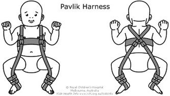
The baby is placed in a soft positioning brace, for 1 to 2 months to keep the thighbone in the socket. This special brace is designed to hold the hip in the proper position while allowing free movement of the legs and easy nappy changes. The brace helps tighten the ligaments around the hip joint and promotes normal hip socket formation. Parents play an essential role in ensuring the harness is effective.

1 month to 6 months
Similar to newborn treatment, a baby's thighbone is repositioned in the socket using a brace or similar device. This method is usually successful, even with hips that are initially dislocated. How long the baby will require the harness varies. It is usually worn full-time for at least 6 weeks, and then part-time for an additional 6 weeks.
If the hip will not stay in position using a harness, our specialists may try an abduction brace made of firmer material that will keep your baby's legs in position.
In some cases, a closed reduction procedure is required. This involves gently move your baby's thighbone into proper position, and then apply a body cast (spica cast) to hold the bones in place. This procedure is done while the baby is under anaesthesia.
6 months to 2 years
Older babies are also treated with closed reduction and spica casting. In some cases, skin traction is used for a few weeks prior to repositioning the thighbone. Skin traction puts a gentle force on the skin and stretches the soft tissues around the hip for the change in bone positioning. In some cases the femur may need be openly placed back into the socket. In this procedure, an incision is made at the baby's hip that allows the surgeon to clearly see the bones and soft tissues. X-rays are taken during the operation to confirm that the bones are in position. Afterwards, the child is placed in a spica cast to maintain the proper hip position.
Follow-Up
Hip dysplasia and bracing requires careful monitoring. Typically this is done with serial ultrasounds at 6 weekly intervals in the brace, then X rays at the age of 1, 2, and 5yrs.
Outcomes
If diagnosed early and treated successfully, children can develop a normal hip joint and should have no limitation in function. Left untreated, DDH can lead to pain and osteoarthritis by early adulthood. It may produce a difference in leg length or a "duck-like" gait and decreased agility.
Even with appropriate treatment, hip deformity and osteoarthritis may develop later in life. This is especially true when treatment begins after the age of 2 years.
Frequently Asked Questions
My child has hip dysplasia - will they be in pain?
Hip dysplasia in young children is typically non-painful, when children become older >6-8yrs then pain can occur.
How long will my child need their brace for?
This depends upon how they grow and how the hip dysplasia resolves. Typically the minimum time a brace is required is 3 months. However, it is not uncommon for children to require braces up to 12-18months in severe cases.
Will my child have a developmental/walking delay due to being in a brace?
Most children who have been in a brace will have a delay in their ability to walk, but they normally catch up very quickly when the brace is removed.
Can my child walk in the brace?
In a hippo or rhino brace, children can often stand or crab walk. They can also usually pull themselves up to stand.
Can my child do tummy time in the brace?
Tummy time is difficult in any brace or orthotic. When the brace is off, it is recommended to encourage this as much as possible.
What is my child’s risk of arthritis in the future?
If hip dysplasia is detected early and treated successfully, there is a low risk of arthritis, however, if there is residual dysplasia, or if the condition is detected late, this increases the risk of arthritis.